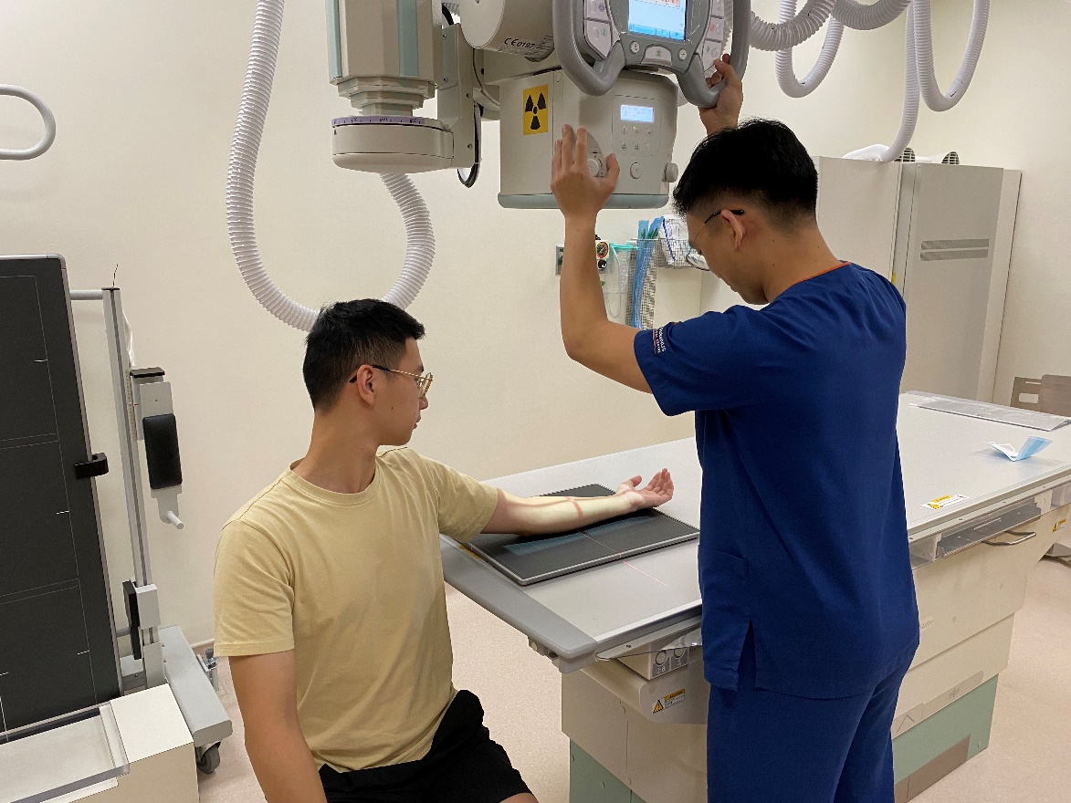
Plain radiography uses X-rays to create 2D images of the human body. A diagnostic radiographer who is trained in imaging the human body will perform the x-ray examination.
A specialist doctor known as a radiologist, will evaluate the images and create a report based on your X-ray images.
Some of the commonly performed examinations performed in SGH include:
- Chest X-ray
Chest x-rays are commonly performed to look for any lung disorders (e.g. lung infections), heart disorders (heart failure) and fractures of the bones in the chest. - Lumbar Spine X-ray
Lumbar spine X-ray is commonly performed to evaluate long-term lower back conditions such as degenerative disc disease. - Knee X-rays
Knee X-rays are commonly performed to evaluate knee conditions such as joint arthritis, joint effusion and fractures.
How to prepare for an X-ray examination?
Preparations can vary for each examination, depending on the region to be imaged. In general:
- Please come in loose and comfortable clothes.
- To allow clear x-ray images to be taken, please remove any jewelry, metallic objects within the region of the x-ray examination.
What happens during an X-ray examination?
The body region will be positioned between the x-ray machine and an imaging plate that creates the x-ray image during the procedure.
Depending on the region, you may have x-rays taken from more than one position to view the region.
It is essential to remain still during the x-ray process as movement can result in blurry images.
Having x-rays taken generally causes no pain. However, there might be discomfort while adopting different body positions. Please inform the staff if you feel pain or discomfort during the process.
What happens after an X-ray examination?
Generally, there are no specific care instructions required after the X-ray examination. The radiographer will send your images to your requesting doctor. Our radiologists will review your image and write a report based on your X-ray images. Both the X-ray image and report will be made available to your primary doctor electronically.
What are the possible risks and limitations for an X-ray examination?
In general, X-ray examinations are very safe and unlikely to produce radiation side effects. The amount of radiation used is very small, so the risks are minimal. Young children and a developing fetus carried by a pregnant woman are more sensitive to X-rays and are at greater risk for tissue damage.
For women of childbearing age, It is important to tell the staff if you are pregnant. X-rays generally are not used on pregnant women because of the possible risk of radiation exposure to the developing baby.
